Athletic injuries are complex because of high velocity and high force impact. Thus, it is always difficult to diagnose and treat such injuries. Traditional MRI’s have limitations in assessing the impact on movement resulting from injury of one or more skeletal tissues.
It is very important to choose the right imaging technique to understand the injury, its type, extent, and location for complete recovery, especially when you need to return to an upcoming sports event. MSK ultrasound imaging is one such technique that is painless, cheap, accurate, and can be done with real-time analysis of a defect or injury.
MSK ultrasound is getting increased popularity because of the safety, cost, and comfort of the patients. Positive outcomes are highly dependent on the correct diagnosis.
NYDNRehab sports medicine clinic has technologies to assess the motion of the joints to treat localized instability and formulate a specific treatment plan. It offers technological advancement to determine biomechanical deviations and is one of its kinds.
MSK ultrasound – Mode of Imaging
An MSK ultrasound uses the same principle of high-frequency sound waves as used in obstetric ultrasound imaging during pregnancy. The difference is that it determines injuries of your musculoskeletal system.
The imaging of ultrasound is not limited to bones and major muscle groups but can also determine injuries of joints and associated tears of the supporting tissue such as tendons and ligaments.
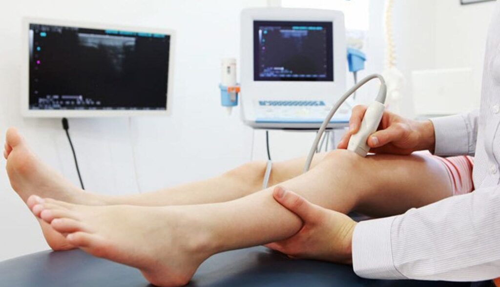
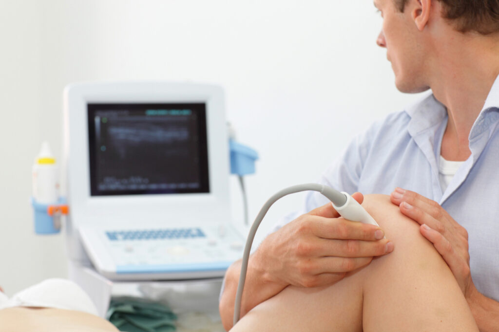
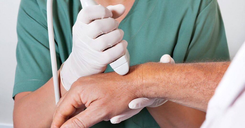
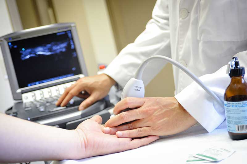
The procedure to perform MSUS consists of these basic steps:
- The gel is applied to the skin of the concerned area of injury.
- A transducer that emits sound waves at high frequencies is then placed on the affected area.
- The waves bounce back from different structures, which are transferred from the probe to create a real-time image of the tissues.
- The difference in density and location of structures creates variations in the echo that is received by the probe and creates different shades on the final image.
Movement of concerning muscle tissue and blood flow to a particular area can be assessed in real-time with this imaging technology.
MSK ultrasound is a great tool to diagnose sports injuries such as tears, bleeding within muscles and joints, inflammation of supporting tissues, early-stage arthritis, fluid accumulation in joints, and tumors of soft tissues.
However, hard-tissue imaging cannot be achieved with MSUS imaging and would warrant a need for X-ray, CT, or MRI.
Advantages of Musculoskeletal Ultrasound Imaging
- It is portable and lightweight equipment that sends sound waves to determine soft tissue injuries.
- It offers real-time detection within minutes after the occurrence of an injury.
- It is better at detecting stress fractures when compared to MRI and X-rays.
- It can even detect injuries to blood vessels, neural bodies, and internal organs with real-time detection and analysis.
- It is safe for use even on pregnant females and can be repeated within a shorter duration because of the absence of harmful radiations or magnetic fields.
- If the MSUS transducer is of high-quality, it can detect even minor injuries such as tears of tendons or the presence of small calcifications and foreign bodies.
- Different variations of MSUS like elastography can predict the extent of activity that can be resumed post-treatment.
- A Doppler ultrasound, on the other hand, can easily detect inflammatory changes well before an MRI and even blood markers.
- Guided movements and patient feedback during MSUS imaging can effectively pinpoint the injury location and the exact source of pain.
- It is comfortable and non-traumatic for the patient.
- Injuries and abnormalities of tendons are best detected with ultrasound imaging as tendons are hyperechoic.
- Ultrasound imaging is also used to perform accurate injections and aspirations.
- It is easy to view bilateral structures in one short session.
- Nerve compressions and long muscular injuries can be easily detected by allowing the transducer to follow a certain pathway.
- It is cheaper and requires lesser space and arrangements to conduct MSUS imaging over CT scans and MRI’s.
- It is safe to perform in people having metal implants and pacemakers.
- Superficial structures can be seen very clearly and accurately with the ultrasound technique.
Accurate diagnosis and timely diagnosis of athletic injuries are paramount for planning corrective treatment and rehabilitation. MSUS is not just used for accurate and real-time diagnosis but also for establishing the extent of recovery post-rehabilitation. This gives the clinician an advantage to assess the runner’s fitness for a safe return to the sports activities.
Dynamic imaging with MSUS
This is the most important aspect of ultrasound imaging for athletic injuries. This is especially important in cases when the pain is only reproduced with a particular movement of the joint.
The patient is asked to reproduce the movement that causes the pain while the clinician evaluates the internal structure with ultrasound imaging to pinpoint the exact location of injury or inflammation.
No other imaging modality provides the same degree of accuracy in such conditions.
MSK ultrasound and its utility in the treatment
The dynamic and immediate visualization by the clinician makes it every effective for treatments and procedures such as:
- Giving specialized soft tissue and joint injections.
- Accurate insertion of medication in the desired location.
- Diagnostic aspiration of soft tissue and synovial fluid.
- Assessing inflammation, blood flow, and tissue organization post-treatment to measure the extent of healing.
The clinician is able to see the progression of the needle instantly and can change the approach immediately. This can prevent any unintentional damage to nerves and blood vessels in the way.
It is specifically used to inject medicines in the shoulder and glenohumeral joints with maximum accuracy. This reduces the chance of re-injecting the sites and the need for more advanced and expensive imaging techniques.
Your doctor is the best judge in determining the diagnostic modality needed for determining your injury or condition. Correct and real-time diagnosis is important to provide early and accurate care to the patient so that they can recover and get back to normal motor functioning in a shorter time span.